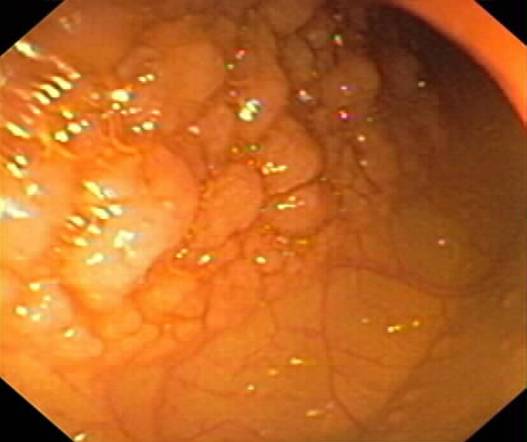
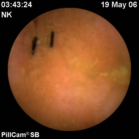
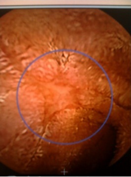
Normal Terminal Ileum
Endoscopic photographs of normal terminal ileum.
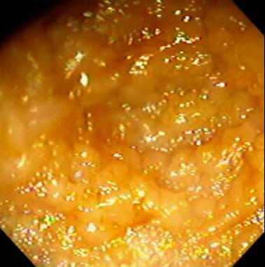
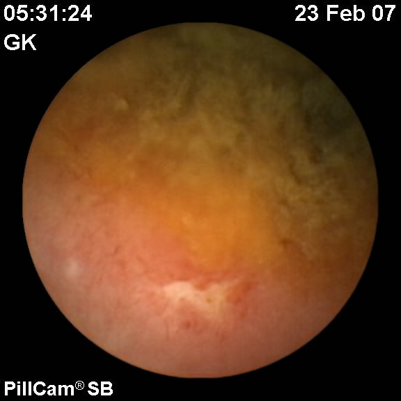
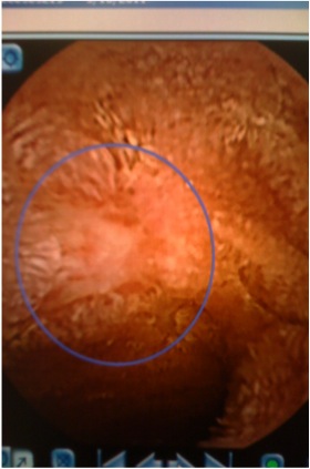
Normal Terminal Ileum
Endoscopic photographs of normal terminal ileum
Normal Terminal Ileum
Endoscopic photographs of normal terminal ileum.

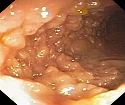
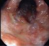
Crohn’s disease
Endoscopic appearance of a 9 yo patient with Crohn’s disease of the terminal ileum.
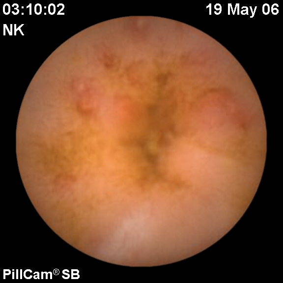
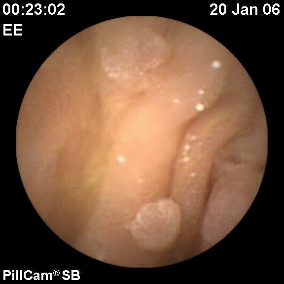
Small bowel apthous lesions
Wireless capsule endoscopy of 14 year old female with Crohn’s disease

Status post-colectomy
Wireless capsule endoscopy of 14 year old female with inflammatory bowel disease, sutures in the pouch
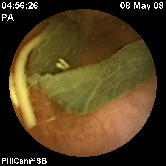
Vegetable matter in lumen
Wireless capsule endoscopy of 15 year old female with chronic abdominal pain

Small bowel ulceration
Wireless capsule endoscopy of 16 year old female with Crohn’s disease

Jejunal polyps
Wireless capsule endoscopy of 18 year old male with familial adenomatous polyposis

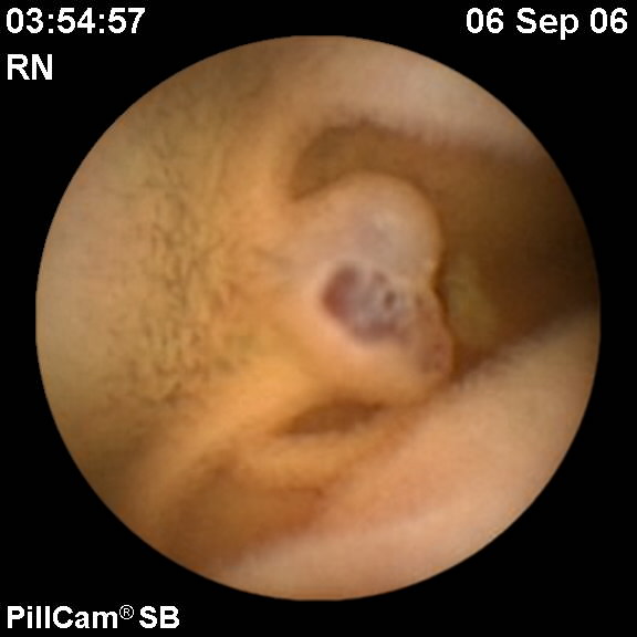
Jejunal venous malformation
Wireless capsule endoscopy of 4 year old female with blue rubber bleb nevus syndrome

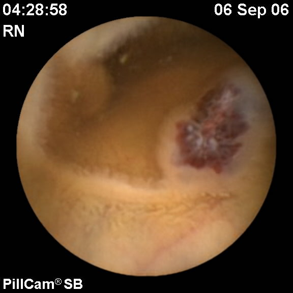
Venous Malformation
Wireless capsule endoscopy of 4 year old female with blue rubber bleb nevus syndrome
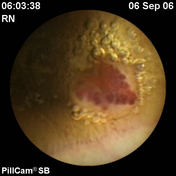
Venous Malformation
Wireless capsule endoscopy of 4 year old female with blue rubber bleb nevus syndrome
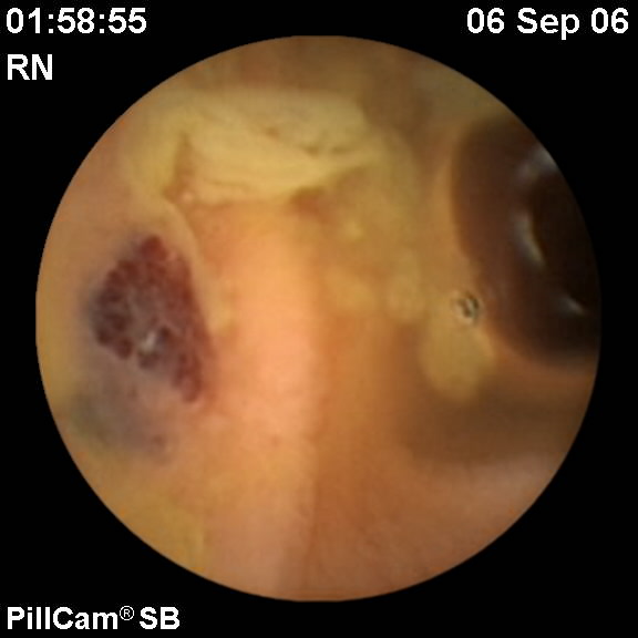
Venous Malformation
Wireless capsule endoscopy of 4 year old female with blue rubber bleb nevus syndrome
Venous Malformation
Wireless capsule endoscopy of 4 year old female with blue rubber bleb nevus syndrome

Capsule Endoscopy
10 year old with acute onset abdominal pain. Capsule endoscopy revealed multiple stellate and linear ulcers throughout the jejunum and ileum

Capsule Endoscopy – Ulcers
Capsule Endoscopy – Ulcers
.JPG)
Crohn’s Disease
Strictures on mid-distal jejunum by double balloon enteroscopy

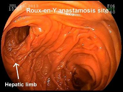
Rouy en y pic
Rouy-en-Y pic Courtesy of Quin Liu MD, Children’s Hospital of Los Angeles
Photos 1-18 (of 18)

.jpg)
.jpg)


.jpg)
.jpg)
.jpg)
.JPG)
Photos 1-18 (of 18)