-

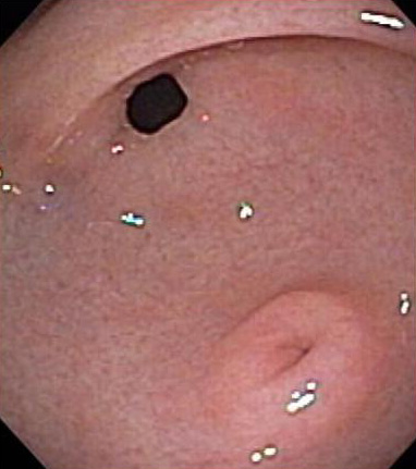
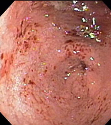
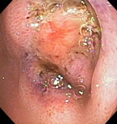
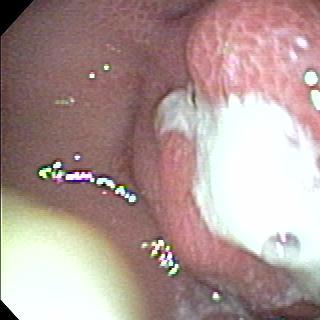
Henoch-Schoenlein Purpura Vasculitis
Endoscopic photograph of the gastric antrum in a 9 yo with vomiting and abdominal pain.
-

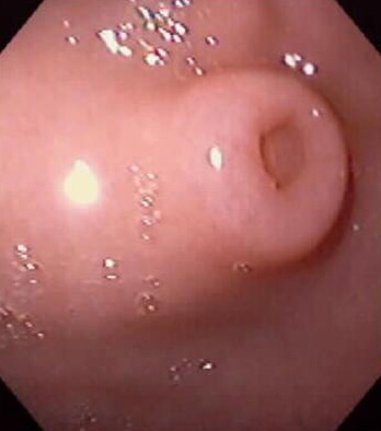
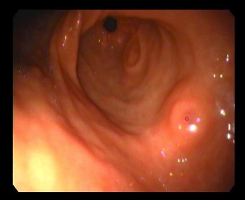
Pancreatic Rest
Endoscopic photograph of a “dimpled” antral mass in a 5 yo found incidentally.
-
Pancreatic Rest
Endoscopic photograph of a “dimpled” antral mass in an 8 yo found incidentally.
-
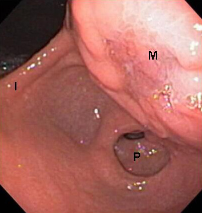
Pancreatic Pseudocyst
Endoscopic photograph of a large pancreatic pseudocyst (M) protruding into the gastric cavity.
-

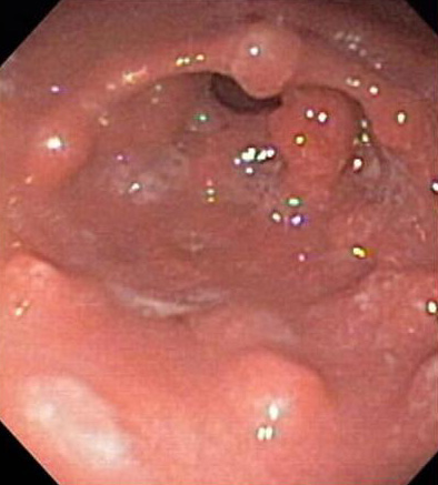
Crohn’s Disease
Endoscopic photograph of a gastric antrum with multiple small nodules in a 16 yo with Crohn’s disease.
-

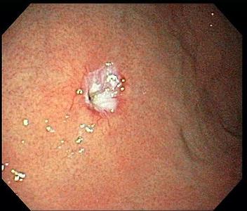
Ulcers
Endoscopic photograph of two gastric ulcers.
-
Normal Fundus
Endoscopic photograph of a normal gastric fundus.
-
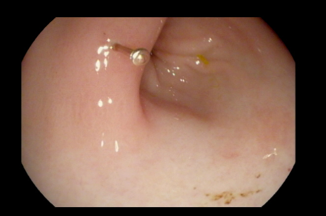
Nissen Fundoplication Defect
Endoscopic photograph of the stomach fundus demonstrating the typical endoscopic appearance after a Nissen Fundoplication has been performed.
-
Gastritis
Endoscopic photograph of severe antral gastritis in a 7 yo who presented with hemetemesis and epigastric pain after developing a viral illness.
-

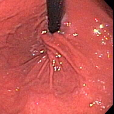
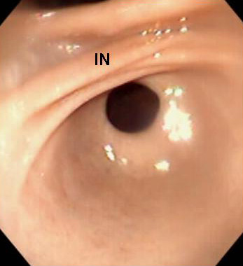
Normal Pylorus
Endoscopic photograph of a normal gastric antrum and incisura (IN).
-

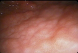
Crohn’s gastritis
An 18 yo with Crohn’s disease underwent surveillance endoscopy.
-
Gastric Fundoplication defect
7 yo male with static encephalopathy.
-
Eosinophilic gastritis
10 yo female with eosinophilic gastroenteritis underwent endoscopy secondary to hypoalbuminemia.
-
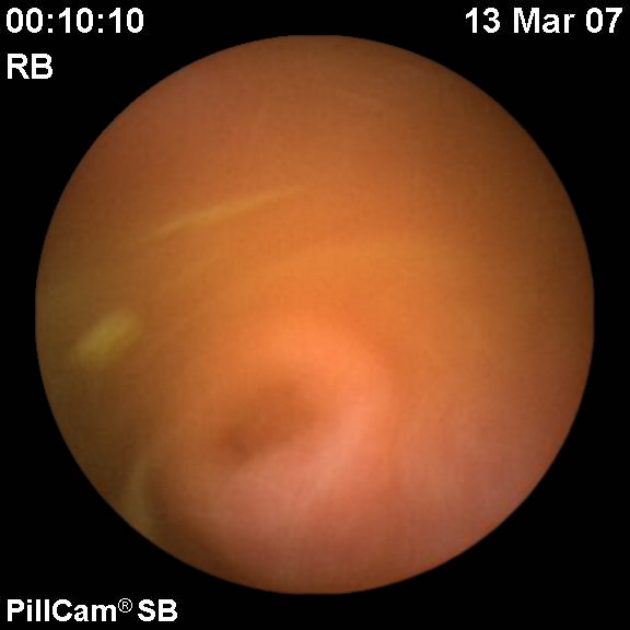
Pancreatic rest
Wireless capsule endoscopy of 18 year old male with inflammatory bowel disease
-

H pylori gastropathy
H pylori gastropathy
-
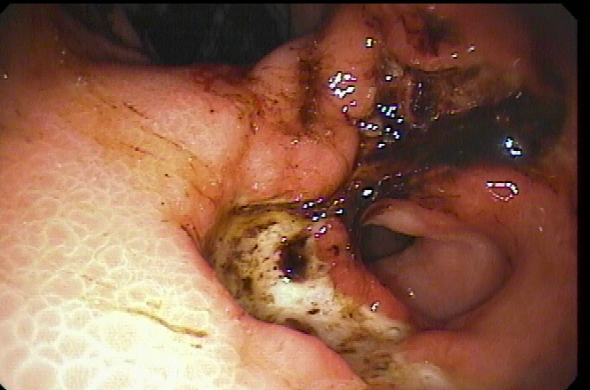
Large Penetrating Gastric Ulcer
Large Penetrating Gastric Ulcer
-
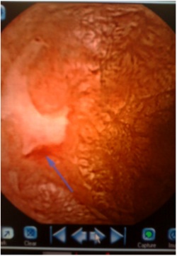
Gastric Polyps
Large penetrating gastric ulcer in patient with Gardner’s syndrome
-
Pin in Stomach
Pin in Stomach
-
Gastric Ulcer
Gastric ulcer in a 19 month-old with chromosomal abnormality presented with melanotic stools and coffee ground gastric contents. Ulcer secondary to G-tube irritation.
-

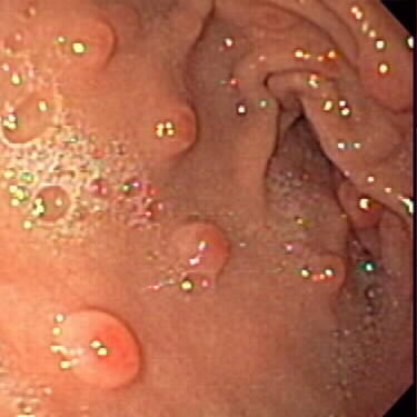
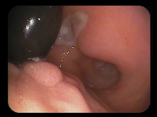
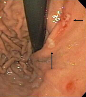
Pancreatic Rests in Antrum
11 year old s/p esophageal atresia repair with reflux. Two raised mucosal lesions with central umbilicated surface consistent with pancreatic rests.
-

Endoscopic clipping of peristsent gastrocutaneous fistula
Endoscopic clipping of peristsent gastrocutaneous fistula Courtesy of Michael Wilsey MD, University of South Florida College of Medicine
-

Burried Bumper Syndrome
Burried Bumper syndrome with drainage from the buried bumper, s/p guide-wire placement to maintain the lumen, s/p traction removal of the PEG tube bumper and successful placement of a gastrostomy button over the guidewire into the gastric lumen. Courtesy of Michael Wilsey MD, University of South Florida School of Medicine
Photos 1-22 (of 22)
.jpg)


.jpg)


Photos 1-22 (of 22)